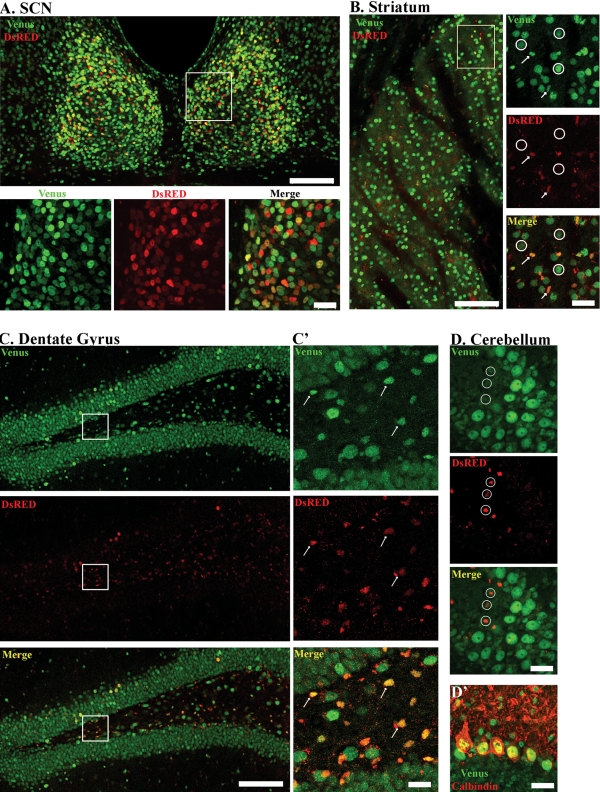
Figure 6.
Brain-specific colocalization of mPer1-Venus and mPer2-DsRED in double transgenic mice. Expression of mPer1-Venus and mPer2-DsRED in the (A) suprachiasmatic nuclei, (B) striatum, (C) dentate gyrus and (D) cerebellum. Boxed regions (in A and B) are provided as higher magnification images in the bottom and right-most panels, respectively. Low magnifications images: scale bar = 100 µm. High magnification images: scale bar = 20 µm. Arrows indicate co-localized expression of Venus and DsRED, whereas circles indicate lack of co-localization. Panel D' represents double-labeling for Venus and Calbindin in the cerebellum. Note that DsRED is not expressed in Purkinje cells of the cerebellum. All animals were sacrificed between ZT 11and 12 and images were captured by confocal microscopy.

