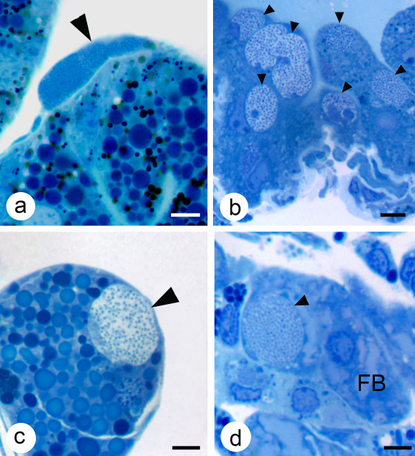
Figure 2.
Light photomicrographs of colonies of A. marginale in various tissues of male D. variabilis. (a) A colony of A. marginale in a gut muscle cell (large arrow); (b) several colonies (small arrows) in Malpighian tubules cells; (c) a colony in a salivary gland cell (large arrow) and (d) a colony of A. marginale (small arrow) in a fat body cell. Mallory's stain, bar = 5 μm.

