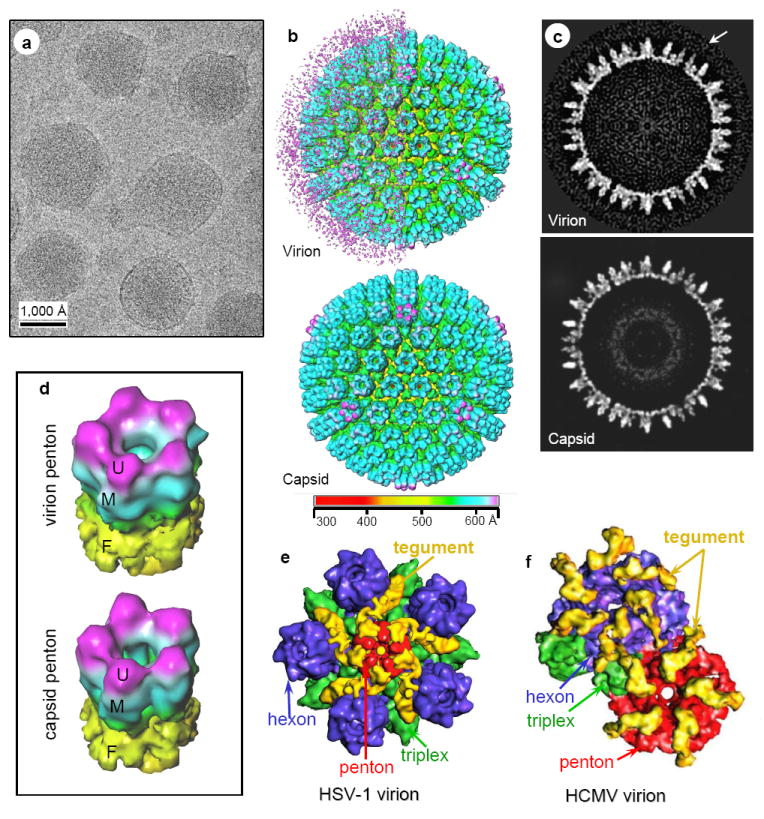
Figure 4. Different inner-layer-tegument/capsid interactions in gammaherpesvirus from those in alpha and betaherpesviruses by single-particle cryoEM averaging.

(A) CryoEM image of MHV-68 virions. (B) Shaded surface representations of MHV-68 reconstructions (30 Å resolutions) viewed along a threefold axis and colored according to radius. Top, Virion reconstruction with left hemisphere displayed at low threshold to show the layer of unordered structure surrounding the capsid. Right hemisphere is displayed at slightly higher density threshold, thus only revealing the enclosed capsid. Bottom, capsid reconstruction. (C) Central slices from the virion (top) and the capsid (bottom) reconstructions, taken perpendicular to a threefold axis. Arrow indicates tegument density layer surrounding capsid surface in the virion. (D) Tilted views of pentons computationally extracted from the virion (top) and capsid (bottom) reconstructions. U, upper domain; M, middle domain; F, floor domain. (E) Close-up view of a computationally extracted portion of the HSV-1 virion reconstruction. The tegument densities, colored in yellow, make contact with the upper domains of penton, and its adjacent triplexes, but not the upper domains of any hexons. (F) Close-up view of a computationally isolated portion of the HCMV virion reconstruction. The tegument densities interact with the upper domains of all penton, hexon and triplex by attaching one end to the upper domain of a hexon (or penton), and anchoring the other end on the upper domain of the adjacent triplex. Color codes in (E) and (F): red, penton; blue, hexon; green, triplex; yellow, tegument.
