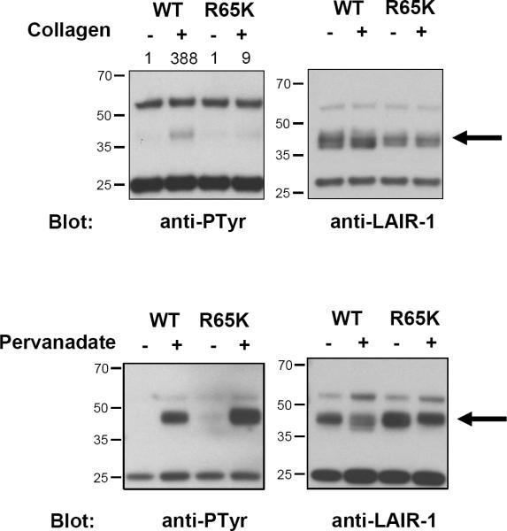
Figure 2.
Tyrosine phosphorylation of LAIR-1 wt and LAIR-1 R65K in transfected K562 cells after interaction with collagen type I (upper panel) or with pervanadate treatment (lower panel). LAIR-1 wt and LAIR-1 R65K were immunoprecipitated after stimulation with clone DX26 antibody. The amount of immunoprecipitated LAIR-1 in each condition was detected by western blot using clone 14 antibody. Numbers on top of the anti-phosphotyrosine blot in the upper panel indicate the relative intensity of phosphorylated LAIR-1. Results shown here are representative of two experiments.

