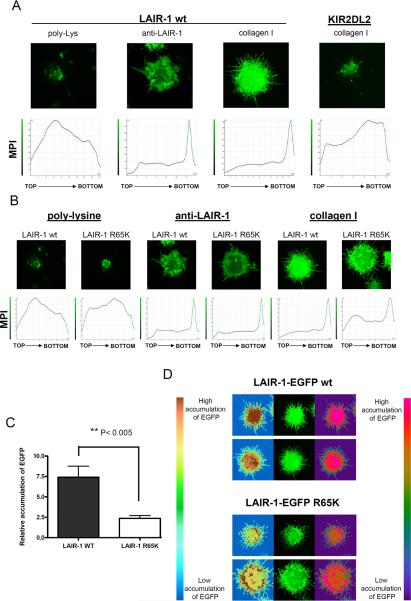
Figure 3.
LAIR-1-EGFP wt polarizes more efficiently than LAIR-1-EGFP R65K to sites of contact with collagen. A) K562 cells transfected with LAIR-1-EGFP wt or KIR2DL2-EGFP were deposited on glass coverslips coated with poly-lysine, anti-LAIR-1 mAb or collagen type I. B) K562 cells transfected with LAIR-1-EGFP wt or LAIR-1-EGFP R65K were deposited on glass coverslips coated with poly-lysine, anti-LAIR-1 mAb or collagen type I. For A) and B), the lower panels show the mean pixel intensity (MPI) of sections of the cell from the top (the distal part of the cell in relation to the glass coverslip) to the bottom (the part of cell in contact with the glass coverslip) and the upper panels show the fluorescence for the section of the cell interacting with the glass coverslip (bottom in lower panels). C) Relative accumulation of LAIR-1-EGFP wt and LAIR-1-EGFP R65K at the site of contact with collagen type I. MPI at the site of contact with collagen was divided by the average MPI of five sections from the middle part of the cell (N= 17). D) Pattern of LAIR-1-EGFP wt and LAIR-1-EGFP R65K polarization is shown in two different false color scales.

