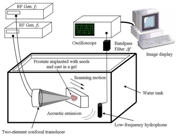
Figure 1.
Experimental VA system diagram. The excised prostate gland implanted with BT seeds is cast in a gel and placed within a water tank at the focus of the confocal ultrasound transducer and scanned at a desired depth inside the sample. The two ultrasound beams differ in frequency by Δf. The hydrophone receives the acoustic emission signal (at Δf) from the prostate. This signal is processed and mapped into an image. [Modified with permission from Ref. 27]

