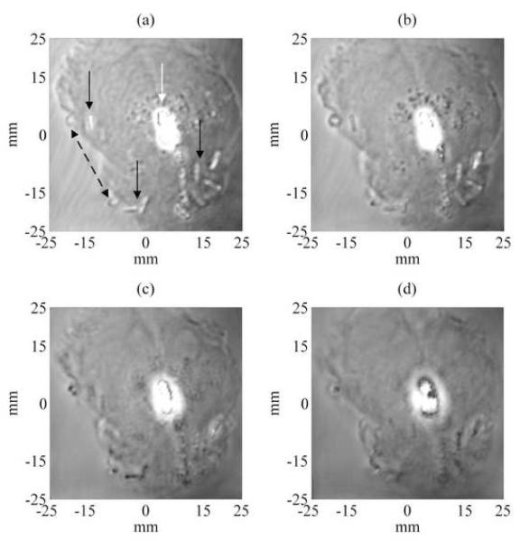
Figure 3.
Experimental magnitude VA images of the excised prostate showing seed location for 4 different depths at 1, 5, 10 and 15 mm (Figs. (a)-(d) respectively) deep from the surface of the prostate gland. The images covered an area of 50 mm by 50 mm, scanned at 0.25 mm/pixel incremental step. These VA images show some of the seeds (pointed by continued black arrows in (a)) at various angles and other seeds appeared more clearly at different depths. One notices also that the intra-prostatic calcifications (pointed by a white arrow in (a)) developed near the center of the prostate gland reflect ultrasound waves and thus obscure some of the seeds. The dotted black double-arrow points to gas bubbles that were developed at the interface prostatic tissue-gelatin after embedding the prostate in the gel phantom.

