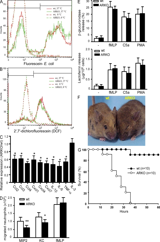
Figure 8.
Neutrophil function and host defense against infection in the absence of AR. (A) Uptake of fluorescein-labeled E. coli by WT and ARKO neutrophils at 37°C. (B) Flow cytometric analysis of oxidant production and subsequent 2′,7′-dichlorofluorescein generation in WT and ARKO granulocytes after PMA stimulation. (C) Relative mRNA expression of chemokines and cytokines in isolated WT and ARKO bone marrow granulocytes. *, P < 0.05. (D) Mature bone marrow neutrophils from WT or ARKO mice were tested for migration in Transwell chambers toward the indicated chemoattractant (200 ng/ml MIP-2, 1 µg/ml KC, or 100 µM fMLP). The number of cells migrated through the Transwell was determined by cell counting. Data from six independent experiments are presented and the error bars represent the SD. *, P < 0.05; **, P < 0.01. (E) Neutrophil degranulation of WT and ARKO neutrophils was examined without or with stimulation by fMLP, C5a, or PMA. (top) The release of the primary granules was examined using the β-glucuronidase activity in the supernatant of stimulated neutrophils. Data are presented as the percentage of total cellular β-glucuronidase content. (bottom) Release of the specific granule marker lactoferrin in the supernatant of stimulated neutrophils was quantified by ELISA. Both degranulation assays were performed in six independent experiments. (F) Representative photograph of WT and ARKO mice after 12-h intraperitoneal injection with E. coli. ARKO mice showed signs of severe infection, such as hunchback, ruffled fur, and eye infection; however, WT mice remained normal. (G) Kaplan-Meier survival curve of WT and ARKO mice in response to intraperitoneal challenge with E. coli. We examined the time course of survival following E. coli infection. Survival over time is shown as percentage mice remaining.

