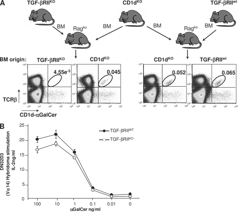
Figure 1.
Functional thymic environment to select iNKT cells in the absence of TGF-β signaling. (A) Flow cytometric analysis of splenocytes in mixed BM chimeras. T cell–depleted BM from 2-wk-old CD4-Cre x TGF-βRIIfl/fl mice or wild-type littermate mice were mixed with BM from CD1dKO mice (ratio 1:1) and transferred into rag2KO animals. 6–7 wk later, the presence in the spleen of iNKT cells was investigated using CD1d-αGalCer tetramers and TCR-β staining. The results are representative of three different experiments with three animals per group. (B) Proliferation analysis of CD1d-restricted Vα14-expressing DN32D3 hybridoma in the presence of thymocytes from either wild-type animals or CD4-Cre x TGF-βRIIfl/fl mice and different concentration of αGalCer. Mean ± SD. n = 2, from two independent experiments.

