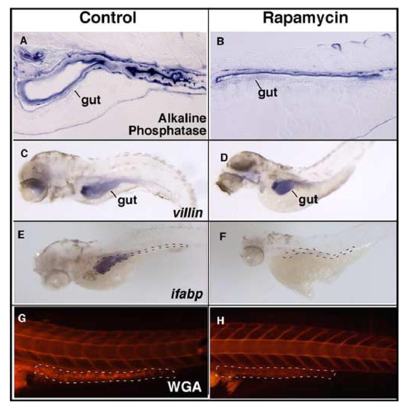
Fig. 5. Effect of rapamycin on intestinal cell differentiation.

(A and B) Histochemical staining for alkaline phosphatase shows enzyme expression and localization to the apical aspect of the epithelial cells. Expression appears to be decreased in the treated embryos, particularly on the ventral epithelium. (C, D) Whole-mount in situ hybridization for the enterocyte marker villin is specific to the intestinal epithelium and is similar in treated and untreated embryos. (E, F) Whole-mount in situ hybridization for the enterocyte marker ifabp shows robust expression in controls, and barely detectable expression in rapamycin treated embryos. (G, H) Lateral view of whole mount stained embryos for goblet cell mucin using rhodamine-conjugated wheat germ agglutinin (WGA). Staining appears to be virtually undetectable in the posterior gut of treated embryos. Gut is outlined by black and white lines (in panels F and H, respectively). Original magnification is 100x in A and B, 40x in C–H.
