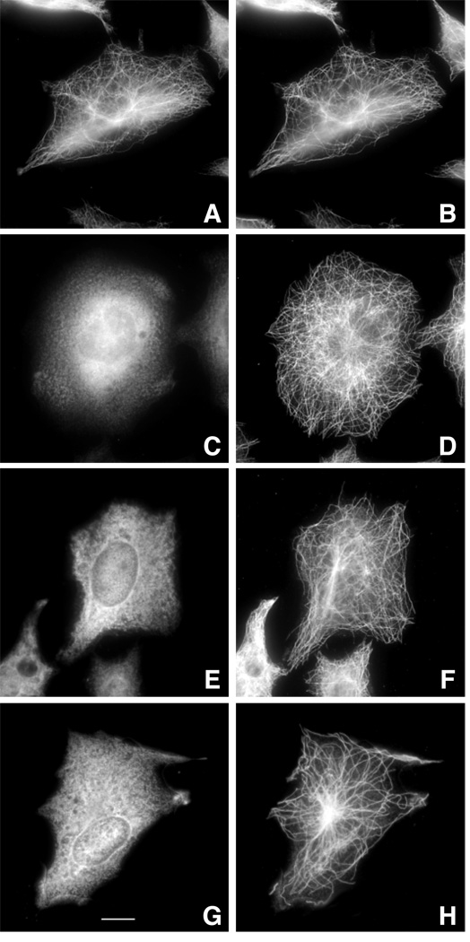
Fig. 2.
Immunofluorescence of transfected cells. Wild-type CHO cells were transfected with wild-type HAβ1-tubulin cDNA (A, B) or with the same DNA containing the 12 amino acid deletion from revertant 6H2 (C, D), the L187R mutation from revertant A5 (E, F), or the Y398C mutation from revertant 5L1 (G, H). At 24 h post-transfection, the cells were fixed with methanol and stained with antibodies to the HA tag (A, C, E, G) and α-tubulin (B, D, F, H). Assembly defective mutant 6H3 was not analyzed in this figure and some of the subsequent figures because we do not know the exact sequence of the truncated gene and therefore cannot construct the appropriate plasmid. The bar in panel G represents 10 µm.

