
Figure 6.
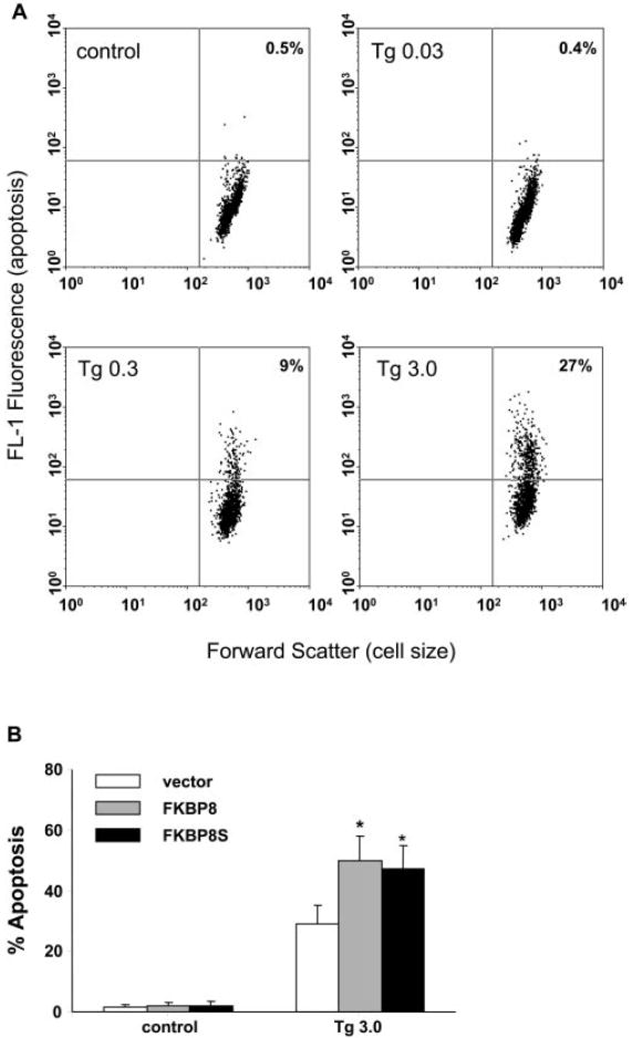
Thapsigargin-induced apoptosis in cultured ARPE-19 cells. (A) ARPE cells stably overexpressing FKBP8 were treated with 0.03 (Tg0.03), 0.3 (Tg 0.3), and 3 (Tg 3.0) μM thapsigargin for 72 hours. Apoptotic cells were stained by TUNEL and analyzed by flow cytometry. The populations of cells in the upper right quadrants were apoptotic. (B) Apoptosis in ARPE cells transduced with vector, FKBP8 and FKBP8s after exposure to thapsigargin for 72 hours. Data represented are the averages of three separate experiments (mean ± SE). *P < 0.05, significant differences from vector, determined by one-way ANOVA and Dunnett multiple comparison test.