
Figure 6. Elevated Akt activity contributes to stabilization of Mcl-1L and c-FLIPL.
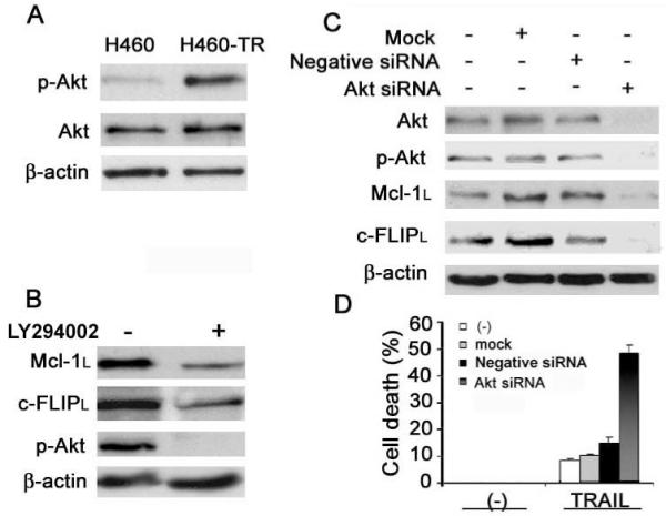
A, Phosphorylated-Akt (p-Akt) and total Akt in H460 and H460-TR cells were measured by Western blot. β-actin was detected as an input control. B, H460-TR cells were treated with 10 μM LY294002 overnight. Mcl-1L, c-FLIPL, and p-Akt were detected by Western blot. β-actin was detected as an input control. C, Pooled siRNA against Akt1, Akt2, and Akt3 was used to knock down Akt in H460-TR cells. p-Akt, total Akt, Mcl-1L, and c-FLIPL were measured by Western blot. D, Nontransfected, mock, and negative siRNA-transfected cells are shown as in H460-TR cells. Control cells were treated with 80 ng/mL TRAIL for 24 h and cell death was detected by LDH assay. Columns, mean of three experiments; bars, SD.