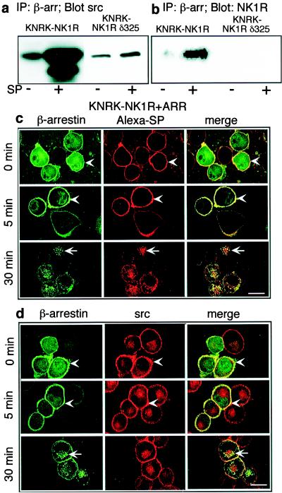
Figure 2.
(a and b) Association of β-arrestin with src and NK1R. KNRK-NK1R and KNRK-NK1Rδ325 cells were treated with or without 10 nM SP for 5 min, crosslinked with DSP, and immunoprecipitated with β-arrestin (β-arr) antibody, followed by Western blotting with src (a) or NK1R (b) antibodies. (c and d) Localization of β-arrestin, src, and NK1R. KNRK-NK1R + ARR cells were incubated with 100 nM Alexa-SP (c) or 10 nM SP (d) for 60 min at 4°C, washed, and incubated at 37°C for 0–30 min. NK1R was localized by Alexa-SP, β-arrestin with GFP, and src by immunofluorescence with a Texas-red-conjugated secondary antibody. Colocalization is shown in yellow in the merged images. Arrowheads indicate plasma membrane, and arrows indicate intracellular localization. (Scale bar = 10 μm.)

