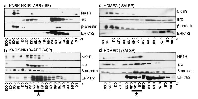
Figure 3.
Gel filtration analysis. Elution profiles from an S300-Sephacryl column of NK1R, src, β-arrestin, and ERK1/2 from KNRK-NK1R + ARR cells (a and b) and HDMEC (c and d). Cells were incubated with (b and d) or without (a and c) 10 nM SP (KNRK cells) or 100 nM [Sar9MetO211]SP (SM-SP, HDMEC) for 5 min, crosslinked with DSP, and fractionated. Western blots of all four proteins in eluted fractions within partition coefficients (σ) of 0.09–0.86 are shown. The region where all four proteins coelute after treatment with NK1R agonists is designated by the star.

