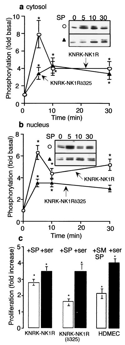
Figure 4.
Nuclear translocation of ERK1/2. (a and b) pERK1/2 in KNRK-NK1R (○) and KNRK-NK1Rδ325 (▴) cells after treatment with 10 nM SP for 0–30 min in cytosolic fractions (a) and nuclear fractions (b). (Inset) pERK Western blots. (c) -Fold increase in [3H]thymidine incorporation into newly synthesized DNA of KNRK-NK1R, KNRK-NK1Rδ325, and HDMEC cells after 48 h with 10 nM SP (KNRK cells), 100 nM [Sar9MetO211]SP (SM-SP, HDMEC), or 20% serum (ser), as compared with untreated cells. *, P < 0.05 as compared with untreated cells, n = 3 (a and b) and 4 (c).

