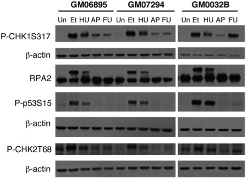
Figure 1.
Activation of DNA damage signaling proteins by FdU treatment. Western blots of extracts from an unaffected cell line, GM06895 and 2 FX cell lines, GM07924 and GM03200B, treated with various DNA damaging agents were probed with the indicated antibodies. Etoposide (Et) was used at 68 µM for 2 h. Hydroxyurea (HU) was used at a concentration of 2 mM for 24 h. Fluorodeoxyuridine, abbreviated in this figure as FU for reasons of space, was used at 1 µM for 18 h. Aphidicolin, indicated here by the abbreviation AP, was used at 0.4 µM for 24 h. Note that the same gel was used for the detection of RPA2 and CHK1 and so the same β-actin control applies. The β-actin data for both experiments is shown beneath the CHK1 data.

