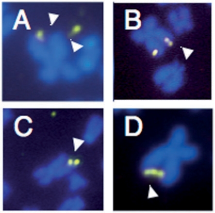
Figure 3.
Representative metaphases seen in FX lymphoblasts after treatment with 1 µM FdU and/or 100 nM UCN-01. (A) X chromosome showing breakage of both sister chromatids. (B) FISH showing X chromosome in which one chromatid shows what appears to be a partial duplication of part of the FRAXA region. (C) X chromosome in which one chromatid has lost the FRAXA region and the other has two copies of that region. (D) X chromosome in which the end of the long arm of the sister chromatids are linked by three copies of the FRAXA region.

