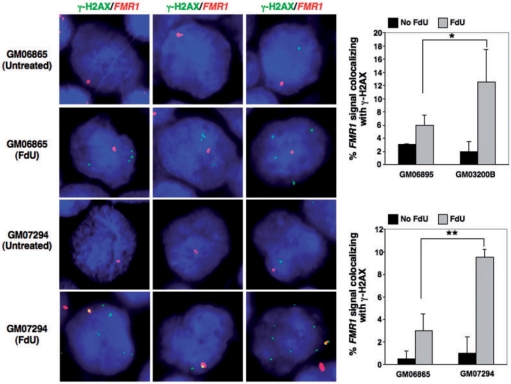
Figure 5.
Colocalization of FX alleles with γ-H2AX foci in the presence of FdU. Immunocytochemistry combined with FISH was used to examine the colocalization of γ-H2AX foci and the FMR1gene in normal (GM06865 and GM06895) and FXS lymphoblastoid cells (GM07294 and GM03200B) treated with or without 1 µM FdU for 18 h. (A) Representative images of γ-H2AX (green) and FMR1 (red) stained cells. (B) Proportion of cells in which γ-H2AX and FMR1 colocalize. The data shows the average of two independent experiments in which a 100 FMR1 signals were counted for each experiment. Error bars signify standard deviation. Statistical analysis was carried out using Fishers exact test. A single asterisk indicates a P-value <0.01, while a double asterisk indicates a P-value <0.005. Note that the X chromosome tends to be located at the nuclear periphery (54). This can make it seem in some cases as if the FMR1 signal is located outside of the nucleus.

