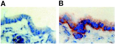
Figure 4.
Indirect immunohistochemistry of t-ASBT in normal rat liver. (A) Liver negative control (no primary antibody was used) displays no staining of cholangiocytes (original magnification ×400). (B) Liver incubated with primary antibody (anti-t-ASBT; 1:50). The basolateral domain of cholangiocytes is decorated (original magnification ×400).

