Figure 1.
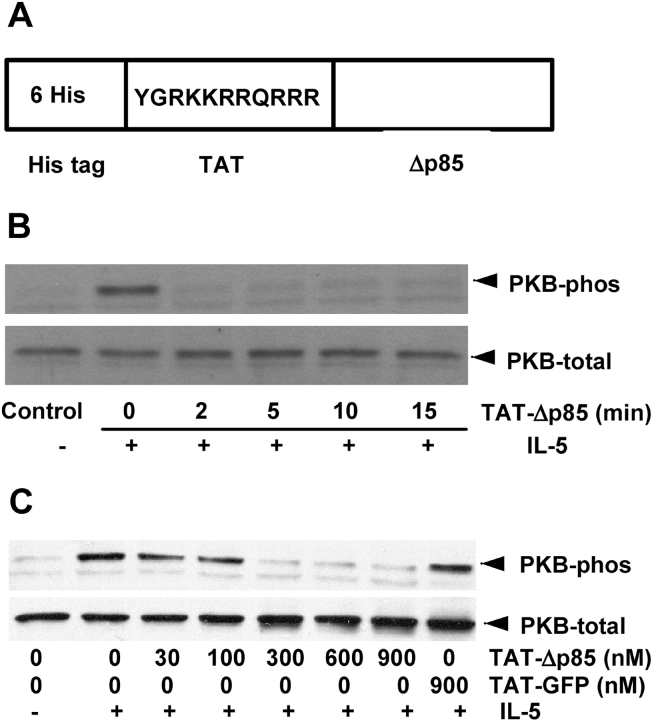
Effect of TAT-Δp85 on IL-5–induced phosphorylation of PKB, a downstream target of PI3K. (A) Structure of TAT fusion protein used in this study. Six His residues and the 11–amino acid TAT peptide precede the N-terminal of the Δp85. The 11 amino acids of TAT are the protein transduction domain. (B) Time-dependent effect of TAT-Δp85 transduction on PI3K activation. Eosinophils were preincubated with 600 nM TAT-Δp85 at 37°C for indicated times and then stimulated with 10 ng/ml IL-5 for 10 min, and cell lysates were separated by SDS-PAGE and probed with anti-phosphorylated PKB antibody (upper panel, PKB-phos) and with antibody for total PKB (lower panel, PKB-total) to demonstrate equal loading in all lanes. (C) Concentration-dependent effect of TAT-Δp85 transduction on PI3K activation. Eosinophils were preincubated with various concentrations of TAT-Δp85 for 15 min and then stimulated with 10 ng/ml IL-5 for 10 min. PKB phosphorylation and expression were measured as previously described.

