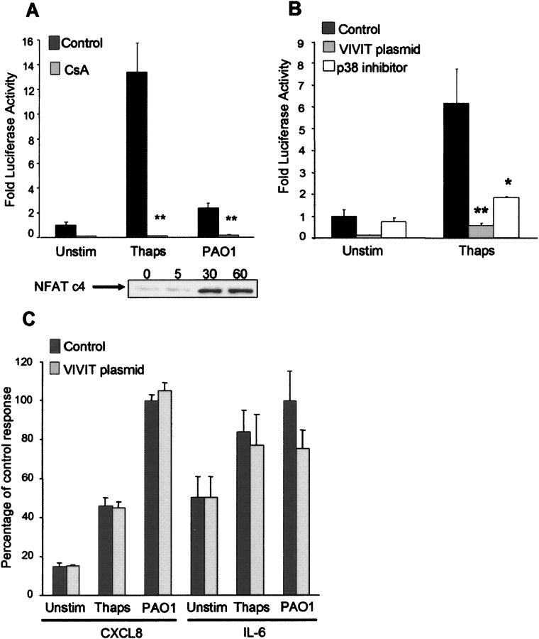
Figure 2.
Epithelial activation of NFAT. (A) 1HAEo− cells transfected with an NFAT luciferase reporter construct and either pretreated for 24 h with 100 nM cyclosporin (CsA) or untreated (Control). Following stimulation for 4 h with 0.1 μM thapsigargin (Thaps) or heat-killed P. aeruginosa PAO1 (108 cfu), luciferase activity was measured and reported as fold activity compared with the unstimulated (Unstim) condition. 1HAEo− cells stimulated with P. aeruginosa PAO1 (108 cfu) were lysed at timed intervals (min) and nuclear lysates screened for the presence of NFATc4. (B, C) 1HAEo− cells transfected with an NFAT luciferase reporter construct and also with the GFP control vector (Control), the expression vector GFP-VIVIT (VIVIT plasmid), or pretreated for 2 h with 6 μM of p38 inhibitor SB202190 (p38 inhibitor). Following stimulation for 4 h with 0.1 μM thapsigargin (Thaps), luciferase activity was measured and reported as fold activity compared with the unstimulated (Unstim) condition. Supernatants were collected and CXCL8 and IL-6 assayed by enzyme-linked immunosorbent assay. Results are normalized to PAO1 (0.49 ng/μg protein ± 0.013 for CXCL8; 2.56 pg/μg protein ± 0.39 for IL-6) indicated as 100% for the purpose of comparison. *P < 0.05, **P < 0.001 compared with control for each stimulus.

