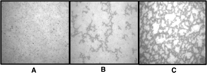
Figure 5.
Epifluorescence microscopy of lung surfactant monolayers reveals a network of interconnected filaments that correlates with two-dimensional gelation measured by the ISR. Lung surfactant monolayers were spread with inclusion of fluorescently-labeled phospholipid, rhodamine DHPE, and examined microscopically after 2 h. (A) Monolayers spread on DMEM media show uniformly bright fields with small, solid-like domains sparsely spread throughout. (B) Monolayers spread on conditioned supernatant from resting A549 AEC show appearances of few dark filaments. (C) Monolayers spread on ROFA-treated A549 cell supernatant show development of a microscopic structural network of highly interconnected dark filaments.

