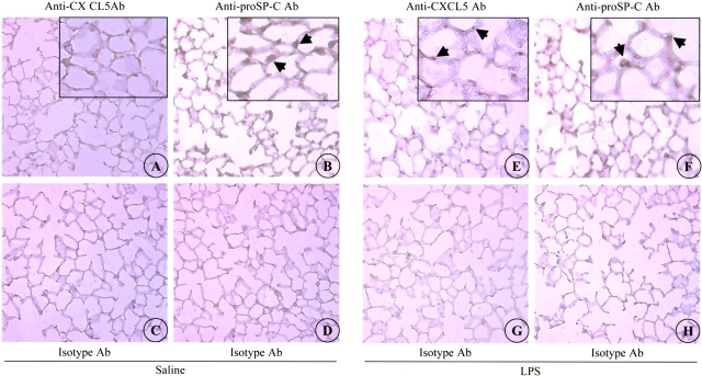
Figure 2.
CXCL5 expression in mouse lung tissue. Immunoreactivity to CXCL5 is shown in the saline-aerosolized lung (A), and the LPS-aerosolized lung (E) at 24 h. Immunoreactivity to the isotype-matched control Ab is shown in the saline-aerosolized lung (C), and the LPS-aerosolized lung (G). Original magnification: ×200. No specific CXCL5 staining was observed in saline-aerosolized lung (A). Alveolar epithelial cells (arrows) are immunostained with the anti-CXCL5 Ab in LPS-aerosolized lung (E). Immunoreactivity to proSP-C was noted in the AEII cells of saline- (B) and LPS-aerosolized lung (F). Images A, B, E, and F are also shown in insets at a higher magnification. No proSP-C staining is observed in sections stained with isotype control Ab (D and H). This is a representative photomicrograph of five independent experiments.

