Figure 4.
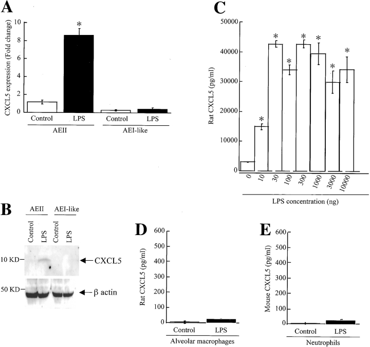
CXCL5 expression by AEII cells in primary culture in response to LPS. Rat AEII cells, AEI-like cells, rat alveolar macrophages, and murine neutrophils were stimulated with 30 ng of LPS. Real-time quantitative PCR was performed in cell lysates 2 h after LPS stimulation or unstimulated (control) and fold change was calculated as described in MATERIALS AND METHODS (A). Cell-associated CXCL5 after stimulation was determined by Western blot (B) and the CXCL5 levels in supernatant was tested 18 h after stimulation (C–E). These results are representative of five separate experiments.

