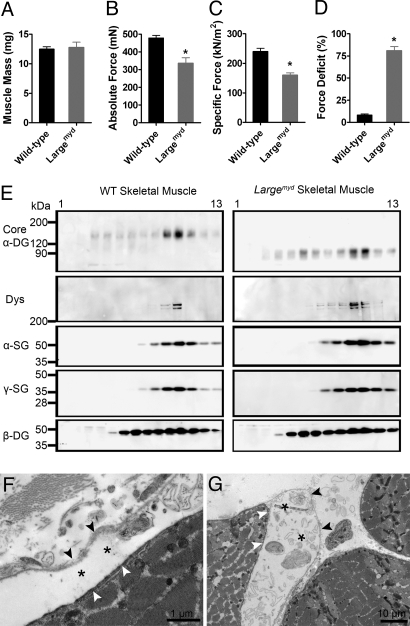
Fig. 3.
Characterization of the contractile properties and the DGC structure in the Largemyd muscle. (A–C) EDL muscle mass (A), maximum force (B), and specific force (C) before subjection to the lengthening-contraction protocol. (D) Force deficits following the lengthening-contraction protocol, as measured for EDL muscles in vitro from C57BL/6 (n = 6) and Largemyd (n = 6) mice. (*, P < 0.05.) All data are presented as the mean ± SEM. (E) Solubilized C57BL/6 and Largemyd skeletal muscle were enriched for DGC by wheat germ agglutinin (WGA) affinity chromatography and separated on 10–30% sucrose gradients. Gradient fractions (1, top; 13, bottom) were blotted with antibodies against core α-DG, dystrophin (Dys), α-sarcoglycan (SG), γ-SG, and β-DG. (F and G) Ultrastructural analysis of quadriceps muscles from Largemyd mice were observed under electron microscopy. Black arrowhead, basal lamina; white arrowhead, sarcolemma; asterisk, dissociation of basal lamina and sarcolemma.

