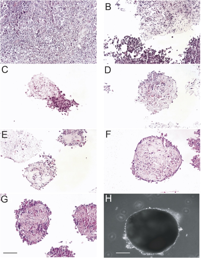
Figure 1.

Tumor fragment spheroids form from human mesothelioma tissue. By hematoxylin and eosin staining, the parental tumor is shown (A), followed by tumor fragment spheroids at Days 1 (B), 3 (C), 5 (D), 7 (E), 10 (F), and 14 (G). Tumor fragment spheroids had consistently formed by Day 10 and showed little change in size during growth in culture. At 2 wk of age, a tumor fragment spheroid is shown with cells around the margin seen as a bright halo (H). Bar, 50 μm.
