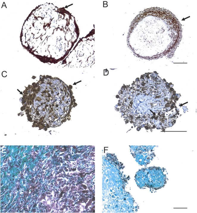
Figure 2.

Mesothelioma cells and collagen are identified in tumor fragment spheroids. Mesothelioma cells were identified by immunohistochemical stains for cytokeratin or calretinin. Shown is expression of cytokeratin (arrows) in a 2-wk-old spheroid (A) and a 6-wk-old spheroid (B) from two different tumors. In 2-wk-old tumor fragment spheroids grown from the same tumor, there is expression of cytokeratin (C) and of calretinin (D) (arrows). By Gomori Trichrome staining, blue-green staining identifies the collagen-rich stroma present in both the original mesothelioma tumor (E) and in the spheroids grown from this tumor at Day 14 (F). Bar, 50 μm.
