Figure 3.
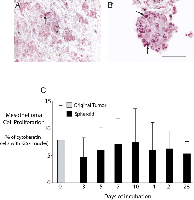
Mesothelioma cells proliferate within tumor fragment spheroids. By double immunochemical staining, cytokeratin-positive cells (pink) could be examined for Ki67 staining (brown) so that proliferation could be determined specifically for mesothelioma cells. Both the original tumor (A) and a 10-d-old tumor fragment spheroid grown from it (B) show Ki67-positive cells (arrows). Bar, 50 μm. (C) In spheroids grown from three tumors, proliferation of mesothelioma cells in the tumor fragment spheroids over 4 wk was compared with that of the original tumor. The proliferation rate was not significantly different from that of the original tumor during the 4 wk. (Mean ± SD; n = 3). Mesothelioma cells continued to proliferate for at least 3 mo (data not shown).

