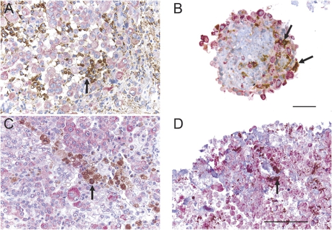
Figure 4.

Macrophages are identified in tumor fragment spheroids. Macrophages, staining with anti-CD68 (brown), were found in the original tumors (A and C) and in tumor fragment spheroids (B and D) grown from those tumors, shown at 2 wk (B) or at 3 mo of age (D) (see arrows). The macrophages could be identified in close proximity to the cytokeratin-positive mesothelioma cells (pink). Bar, 50 μm.
