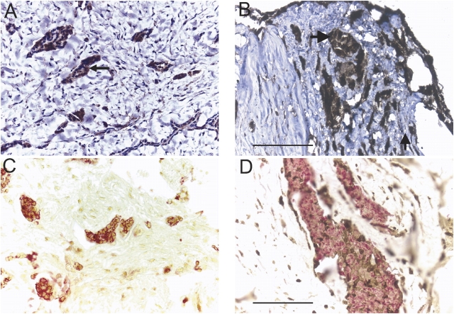
Figure 5.
Mesothelioma cells in tumor fragment spheroids express DR5. By staining for DR5 alone followed by a hematoxylin counterstain, DR5 was shown to be present in clusters of cells (arrow) in both the original tumor (A) and in tumor fragment spheroids at Day 14 (B). By double immunohistochemical staining for DR5 (brown) and cytokeratin (red) without hematoxylin counterstaining, DR5 staining could be localized to the cytokeratin-positive mesothelioma cells in both the original tumor (C) and the tumor fragment spheroid grown from it at Day 14 (D). Bar, 50 μm.

