Figure 1.
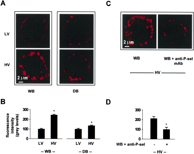
Confocal images of single lung endothelial cells showing P-selectin expression on the cell surface. anti-P-sel mAb, anti–P-selectin monoclonal antibody, RMP-1 (20 μg/ml). HV, high tidal volume; LV, low tidal volume. WB, whole blood perfusion; DB, perfusion with leukocyte- and platelet-depleted blood. The single endothelial cells shown were obtained from lungs treated either under different combinations of ventilation and perfusion conditions as indicated (A, B), or from lungs exposed only to HV ventilation under different perfusion conditions (C, D). In paired experiments, we quantified the mean fluorescence in 30–40 cells from each lung. We processed cells obtained from each lung in parallel but in separate aliquots (n = 3 paired experiments). Mean ± SE. *P < 0.05, compared with bar on left by paired t test.

