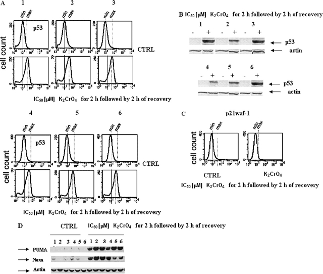
Figure 8.
Effects on p53 protein levels and of target genes (p21waf-1, PUMA and NOXA) in primary cultures of human bronchus epithelium. (A and C) Cells were treated with K2CrO4 at their IC50 values for 2 h and analyzed after 2 h recovery, collected, washed with PBS, and finally resuspended in 500 μl of 4% paraformaldehyde in PBS for fixing. After 15 min at room temperature, 5 ml of 1% BSA in PBS-Tween (BPT) was added, followed by centrifugation at 300 × g for 5 min. Cells were resuspended in 0.2% Triton X-100 in PBS and incubated for 10 min on ice. After addition of 5 ml of BPT and centrifugation, the cells were incubated in primary antibody solution in 5% nonfat dry milk in PBS-Tween (0.05%). PAb 122, for p53 protein detection, and polyclonal anti-p21waf1 antibody (Abcam) were used. All antibody incubations were done in room temperature for 1 h. The cells were washed once with 5 ml BPT and incubated with the appropriate secondary antibody in BPT. After washing, the cells were analyzed in FacsCalibur (BD Biosystems). Data are representative of three replicate experiments yielding similar results. (B–D) In the same pool of cells as that of (A), p53 (B), PUMA and NOXA (D) protein increases were evaluated by Western blotting. Data are representative of three replicate experiments yielding similar results.

