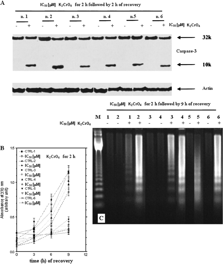
Figure 9.

Induction of caspase-3 and apoptosis in primary human bronchoalveolar cells. (A) Induction of caspase-3 activation and apoptosis in primary cultures of human bronchus epithelium. Cells were treated with μM K2CrO4 at their IC50 values for 2 h and analyzed after 2 h recovery. Data are representative of three replicate experiments yielding similar results. (B) Kinetics of drug-induced apoptotic cell death evaluated by ELISA (Roche Diagnostics) according to manufacturer's instructions (catalog no. 1585 045). A total of 104 cells/well were incubated for 2 h with K2CrO4, their IC50 values, and then cells were incubated in drug-free medium for additional time periods. After the times indicated, 100 μl/well of supernatant plus 100 μl/well of lysates were removed and tested by ELISA. Data are expressed as mean ± SE of two independent experiments performed at least in duplicate and yielding similar results. (C) Internucleosomal DNA fragmentation. Cells were treated as in (A), and after 9 h of recovery, cells, including those floating and adherent, were collected. Data are representative of three replicate experiments yielding similar results.
