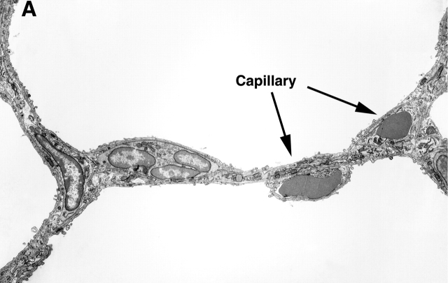
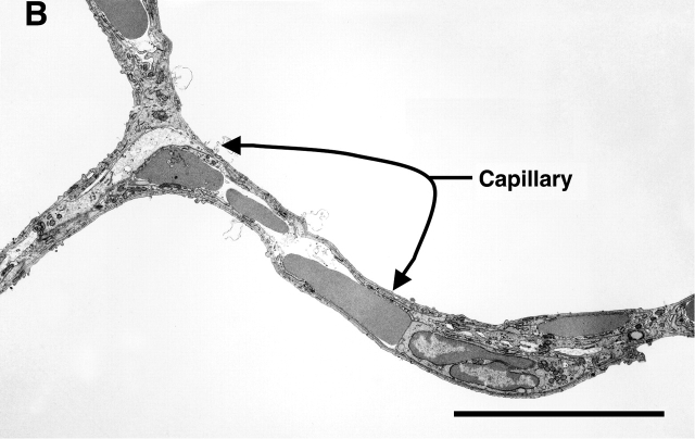
Figure 4.
Representative transmission electron micrographs of the lungs of Day 21 control (A) and SU1498-treated (B) mice. A demonstrates an alveolar wall with normal appearing capillaries that each contain a single red blood cell. In contrast, the alveolar wall of the SU1498-treated mice contains enlarged capillaries with multiple red blood cells demonstrated in the lumen (B). Representative capillaries are indicated by the arrows and labels. Original magnification: ×3,000; bar denotes 10 μm.


