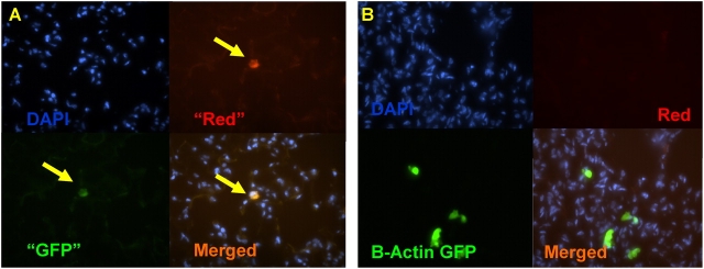
Figure 3.
Distinguishing autofluorescent artifact from engraftment is not possible with dual channel immunofluorescence microscopy. (A) Lung frozen tissue section from a wild-type mouse that has not been exposed to antibody staining shows rare cells that exhibit equal red and green autofluorescence (arrows). (B) Recipient lung after bone marrow transplantation from a B-actin–GFP mouse (no antibody exposure). Donor-derived cells in this section fluoresce green but not red, indicating true GFP+ fluorescence. Nuclei are counterstained with DAPI.

