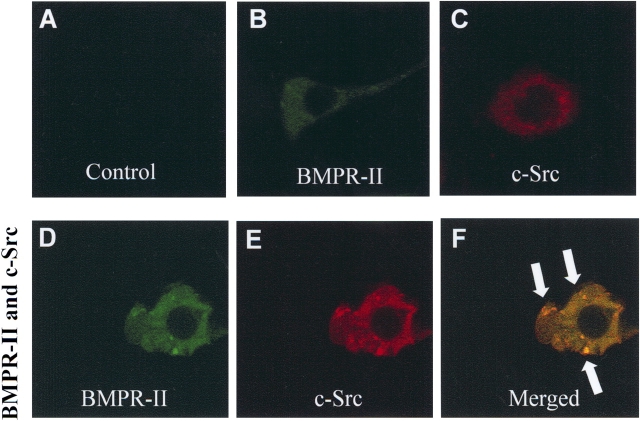
Figure 3.
Recruitment and co-localization of c-Src tyrosine kinase with BMPR-II. (A) Confocal images of negative control (without primary antibody). (B) 6xHIS-BMPR-II expressed alone. HEK293 cells were transfected with wild-type BMPR-II, permeablilized, and stained with antibody against 6xHIS and FITC-conjugate anti-HIS antibody to visualize BMPR-II (green). (C) HA–c-Src tyrosine kinase expressed alone. Cells were detected by using antibody against HA and rhodamine-conjugated monoclonal anti-HA antibody to visualize c-Src tyrosine kinase (red). (D–F) Confocal images of (D) 6xHIS-BMPR-II and (E) HA-c-Src tyrosine kinase in the same HEK293 cell overexpressing both BMPR-II and c-Src tyrosine kinase. (F) 6xHIS-BMPR-II colocalized with HA-c-Src tyrosine kinase in intracellular vesicle-like aggregates (indicated by arrows) in the merged image. Data were representative of three independent experiments.

