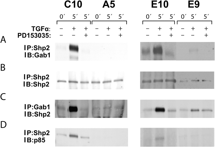
Figure 5.
Complexes containing Shp2 with Gab1 and p85 in C10 and E10 cells. (A and B) C10/A5 and E10/E9 paired nontransformed/malignant cell lines were serum starved and treated with TGF-α with or without EGFR inhibitor PD153035. Lysates were immunoprecipitated with anti-Shp2 and immunoblotted sequentially with anti-Gab1 and anti-SHP2. In the nontransformed cells, the complex was increased in C10 and E10 cells by TGF-α, and this increase was blocked with PD153035. No Shp2/Gab1 containing complex was detectable in A5 cells; a low level was noted in E9 cells. (C) The four cell lines were treated as descibed previously, and lysates were immunoprecipitated with anti-Gab1 and immunoblotted with anti-Shp2. Results confirm the formation of a PD153035-inhibitable complex in the E10 and C10 cells, with much less in the A5 and none in the E9. (D) The four cell lines were treated as described previously, and lysates were immunoprecipitated with anti-Shp2 and immunoblotted with anti-p85. This complex was formed in response to TGF-α and was inhibited by PD153035 in C10 and E10 cells but not E9 and A5 cells.

