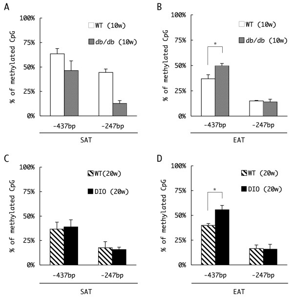
Figure 5.
Comparison of the DNA methylation profile of the peroxisome proliferators activated receptor γ (PPARγ) in white adipose tissue (WAT). Genomic DNA was extracted from subcutaneous adipose tissue (SAT) (a, c) and epididymal adipose tissues (EAT) (b, d) of 10 week-old wild-type (WT) or db/db mice (a, b) or 20 week-old WT/diet-induced obesity mice (c, d). Genomic DNA prepared from each tissue was treated with sodium bisulfite, and amplified by polymerase chain reaction (PCR) with the primers designed for the flanking regions of the -437 bp or -247 bp CpG site. The methylation status of each site was estimated by the efficiency of restriction endonuclease digestion of the PCR amplicon, and the percentages of the methylated fragments are represented (n = 3, mean ± SD, *P < 0.05, t test).

