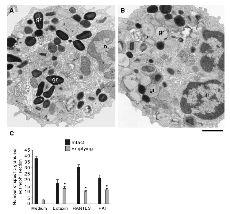
Figure 1. Morphologic effect of three physiologic stimuli on human eosinophil specific granules.

Cells were incubated with control buffer (A, C), 100 ng/mL eotaxin (B, C), 1 μM platelet-activating factor (PAF) (C) or 100 ng/mL regulated on activation, normal, T-cell expressed and secreted (RANTES) (C), immediately fixed and prepared for transmission electron microscopy. After 1 h of stimulation, granules exhibited progressive emptying of their contents and showed morphological diversity. Not all granules exhibited content losses. (C) Significant increases in numbers of emptying granules occurred after stimulation with the three stimuli (*p < 0.05). Eosinophils were isolated by negative selection from healthy donors. Counts were derived from three experiments with a total of 3945 granules counted in 95 electron micrographs randomly taken and showing the entire cell profile and nucleus. gr, granule; n, nucleus. Scale bar, 1.9 μm (A, B).
