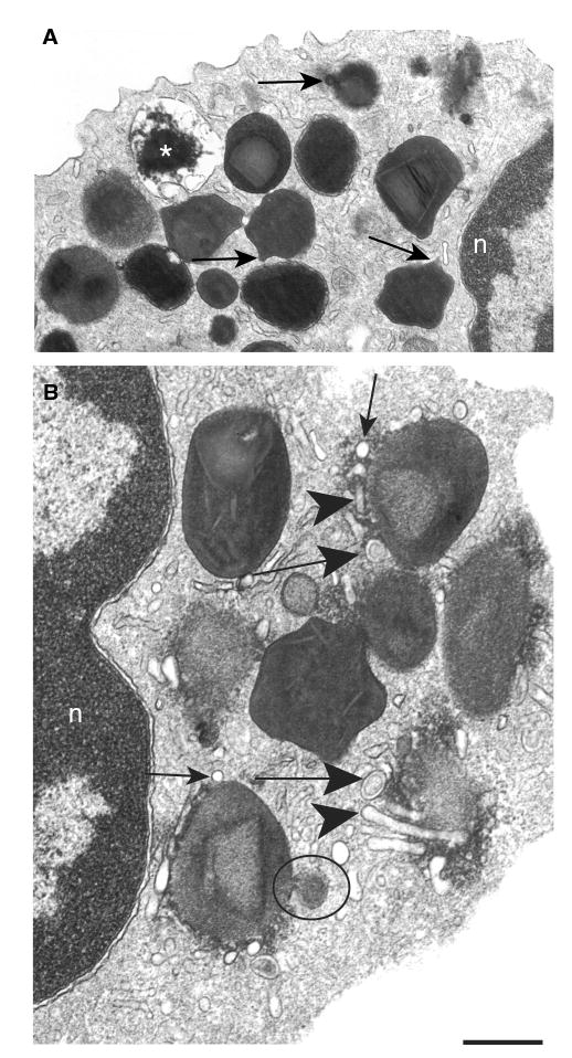
Figure 4. Early stimulus-induced granule events in human eosinophils.

(A) After 30 min of stimulation, specific granules showed ill-defined cores and matrices and became irregular with elongation of their surfaces (arrows). An emptying granule is indicated (*). (B) A prominent system of vesicular compartments composed of small round vesicles (thin arrows), Eosinophil Sombrero Vesicles (EoSVs) (large arrows) and tubules (arrowheads) was seen at the granule surface. A large vesicle profile budding from the granule surface shows the same granule density (circle). Eosinophils were isolated and stimulated with eotaxin as in Figure 1. Transmission electron microscopic evaluations were derived from three experiments, and specimens were studied at magnifications ranging from ×5000 to ×75 000. n, nucleus. Scale bar, 720 nm (A) and 400 nm (B).
