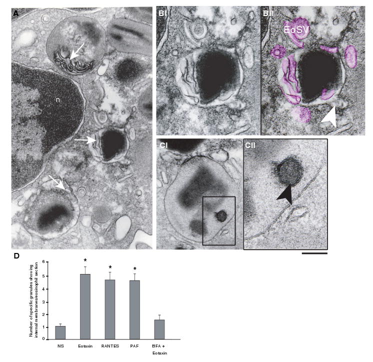
Figure 6. Emptying granules exhibit internal membranous domains in agonist-stimulated eosinophils.

(A) Transmission electron microscopy (TEM) revealed intragranular membranous subcompartments organized as a tubular system in the matrix area (arrows). (B) Higher magnification of an emptying granule with internal membranous tubules (highlighted in pink in Bii) seen in conjunction with mobilized core. Eosinophil Sombrero Vesicles (EoSVs) around the granule are also colored pink. A budding vesicle is indicated (arrowhead). (Ci) A mobilized granule shows part of its content inside a subcompartment (box). (Cii) Higher magnification of the boxed area shows that a true internal membrane (arrowhead), with the same trilaminar appearance as exhibited by the outer granule membrane, encircles part of the granule content. (D) The numbers of granules showing internal membrane domains significantly increased after stimulation (*p < 0.05), but in brefeldin A (BFA)-pretreated eosinophils, these numbers did not differ from unstimulated cells. Cells were stimulated with eotaxin (A, B), regulated on activation, normal, T-cell expressed and secreted (RANTES) (C, D), platelet-activating factor (PAF) (D) or BFA prior to eotaxin (D), processed for TEM and analyzed as in Figure 1. n, nucleus. Scale bar, 480 nm (A), 300 nm (B), 350 nm (Ci) and 138 nm (Cii).
