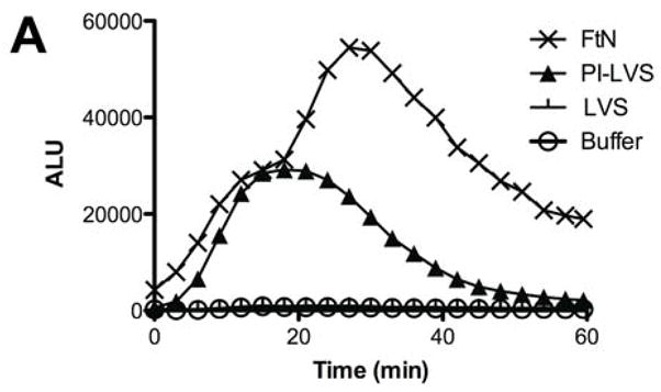
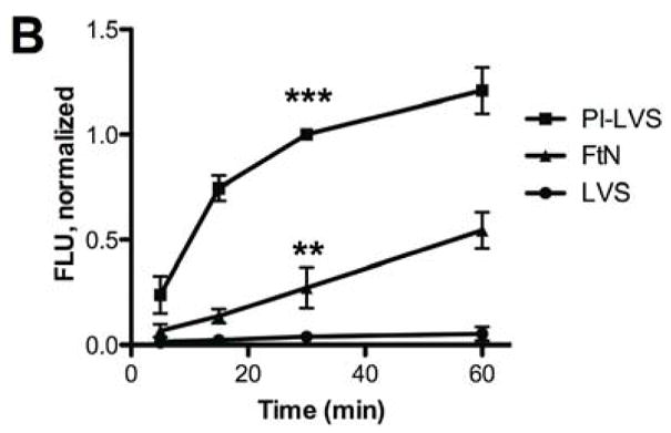
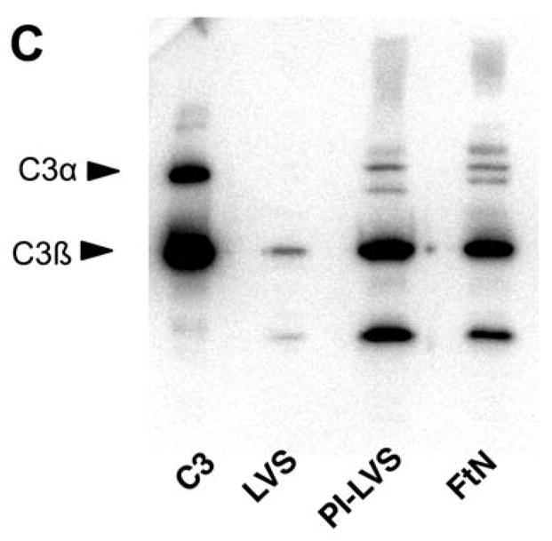
Fig. 1.


FtN stimulated LCL in human PMN and fixed complement component C3. (A) Bacteria were incubated in 50% PHS for 30 min, washed and incubated with human PMN. Control PMN were incubated in buffer without bacteria. Reactive oxidant species production was measured using LCL every 30 seconds. ALU, arbitrary light units. Results are representative of five experiments. (B) Bacteria were incubated in 50% PHS, and opsonization was stopped at the indicated times by adding ice-cold buffer with ethylenediaminetetraacetic acid (final concentration, 10 mM). Bacteria were then washed and incubated with anti-C3 fluorescent antibody. FLU, fluorescence light units. FLU were normalized based on the fluorescence of PI-LVS at 30 min. **, P < 0.01 compared to LVS at 30 min. ***, P < 0.001 compare to LVS at 30 min. N= 3. (C) Bacteria incubated in 50% PHS for 30 min were washed and subjected to immunoblotting for C3. Immunoreactive proteins corresponding to C3α and C3β are marked on the left. The α′-chain of C3 and its fragments can be seen on the blot as higher molecular weight bands that migrate more slowly than the C3 β-chain because they remain covalently bound to their bacterial targets. C3, purified complement component C3. The blot is representative of four independent experiments.
