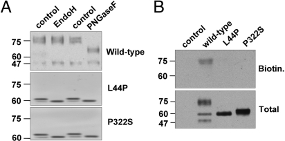
Fig. 2.
Subcellular localization of WT-V2R and V2R mutants in NDI. (A) MDCK WT-V2R, V2R-L44P, or -P322S protein samples were digested with Endoglycosydase H or Protein N-glycosydase F. Subsequently, protein samples and their respective undigested controls were analyzed by immunoblotting. (B) MDCK cells expressing WT-V2R, V2R-L44P, or -P322S were subjected to cell surface biotinylation and equalized total lysate and cell surface samples were analyzed by immunoblotting.

