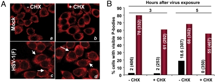
Fig. 3.
HSV-1(F)-induced P-bodies do not disappear after treatment with CHX. HeLa cells grown in 4-well slides were either mock-infected or exposed to 10 pfu of HSV-1(F) per cell for 3 or 5 h. At these times the inoculum was replaced with spent medium alone or medium containing 50 μg of CHX per mL of medium. After 1-h incubation at 37 °C, the cells were fixed and immunostained with antibody against Rck/p54 (red). The cells were counted in 10 randomly chosen fields. The percentages of cells exhibiting visible P bodies were counted and shown as a percentage of total cells counted as indicated in parentheses. (A) Representative images of mock- and HSV-1(F)-infected cells either untreated (a and c) or incubated for 1 h in medium containing 50 μg of CHX per mL 3 h after infection (b and d). (B) Histogram showing the percentage of cells containing visible P-bodies in mock- and HSV-1(F)-infected cells untreated or CHX-treated after either 3 or 5 h of exposure to the virus. Total cells counted are reported in parentheses.

