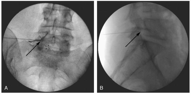
FIGURE 1.
Final needle position was confirmed with the needle tip within the lateral half of the pedicle on AP view and within the foramen on lateral view. Panel A shows the needle tip (arrow) below the pedicle within the intervertebral foramen. Subpedicular dye spread of contrast dye into the epidural space confirms transforaminal placement. The lateral fluoroscopic image shown in B shows the needle tip within the foramen.

