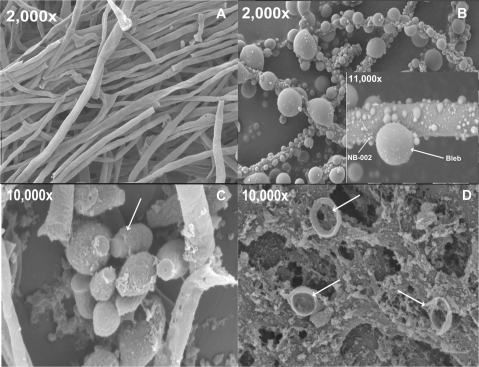
FIG. 2.
Scanning electron micrographs of T. rubrum NBD030 after treatment with NB-002 for 1 h at room temperature. (A) Control mycelia (no treatment or treatment with vehicle); (B) treatment of mycelia with 100 μg NB-002; inset, higher magnification to show the differential size of the nanoemulsion droplets (white arrow, NB-002) compared to the various sizes of the extrusions (blebs); (C) control microconidia (no treatment or treatment with vehicle; white arrow, microconidial spore); (D) treatment of microconidial spores with 12.5 μg NB-002, with white arrows indicating broken spores.

