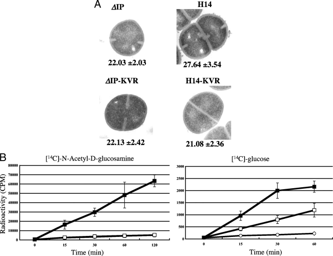
FIG. 1.
(A) Transmission electron micrographs of ΔIP derivative strains. The values given under each panel are the means and standard deviations of the cell wall thicknesses of the cells (in nanometers). Magnifications, ×30,000. (B) Incorporation of [14C]N-acetyl-d-glucosamine or [14C]d-glucose into the cell wall of the N315 derivative strains. Open squares, parent recipient ΔIP; open circles, H14-KVR; closed squares, H14. The counts per minute were measured at the indicated time points. The experiment was performed in triplicate on three independent occasions, and the results are shown as the mean values ± the standard deviations.

