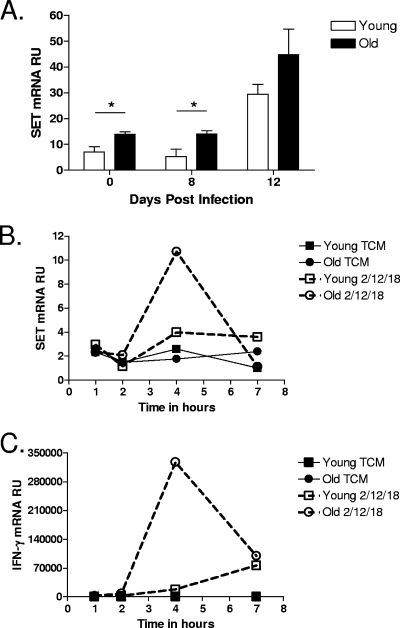
FIG. 6.
The level of SET expression is elevated in CD8 T cells isolated from the lungs of old mice. (A) Young and old mice were infected aerogenically with M. tuberculosis, and lungs were harvested at 0, 8, and 12 days postinfection (n = 4 to 5). Single-cell suspensions were obtained by gentle enzymatic digestion, and CD8 T cells were purified by use of magnetic beads. (B and C) Single-cell suspensions were obtained from the spleens of naïve young and old mice (n = 5), and CD8 T cells were purified by use of magnetic beads and pooled by age. CD8 T cells were cultured with a cytokine cocktail of IL-2 (100 ng/ml), IL-12 (5 ng/ml), and IL-18 (10 ng/ml) for 1, 4, and 7 h. Cells were homogenized in Trizol and frozen at −80°C. RNA was isolated and reverse transcribed, and cDNA was amplified for SET and IFN-γ by RT-PCR. The relative level of expression of SET (A and B) or IFN-γ (C) message is shown as means ± SEM (A) or as relative units (RU) (B and C). Statistical significance between young and old mice at each time point was determined using the Student t test (A) (*, P value of <0.05). TCM, tissue culture media.

