FIG. 6.
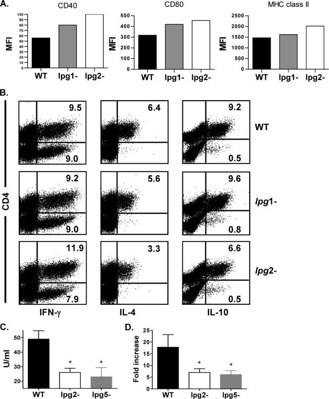
Differences in the early immune response in mice infected with lpg2− L. major are unrelated to absence of LPG. BALB/c mice were infected in the footpad with 5 million WT, lpg1−, and lpg2− L. major stationary-phase promastigotes. After 3 days, mice were sacrificed and CD11c+ cells (DCs) were isolated from pooled dLN cells; stained for surface expression of MHC class II, CD40, and CD80 molecules; and analyzed by flow cytometer (A). Some unfractionated dLN cells were also stimulated with SLA for 3 days, and the percentage of IFN-γ-, IL-4-, and IL-10-producing cells was determined by intracellular cytokine staining (B). In some experiments, dLN cells from mice infected with WT, lpg2−, and lpg5A− lpg5B− L. major for 3 days were cultured for 72 h, and the culture supernatant fluid was assayed for IL-4 by ELSA (C). Total cellular RNA was isolated from some dLN cells and quantified by real-time PCR (D). Data presented are representative of two independent experiments (n = 3 to 5 mice/group) with similar results. *, P < 0.05.

